Respiratory Auscultation: A Comprehensive Guide to Breath Sounds
Listening to breath sounds, respiratory auscultation, is a crucial clinical method for assessing respiratory problems in patients. This article provides detailed descriptions of different respiratory sounds, accompanied by audio recordings for educational purposes.
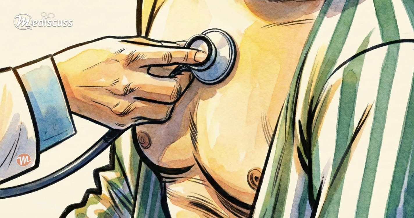
I encourage you to listen to all the audio samples on this page. For an experience similar to using a stethoscope, it is advisable to use high-quality headphones or earphones.
Note on Equipment: Most of the breath sounds in this article were recorded using a Littmann 3200 electronic stethoscope, and some using the Littmann CORE digital stethoscope that I currently use. These are widely respected electronic stethoscopes for auscultation.
Auscultation Basics
To effectively perform auscultation, certain conditions and practices should be observed to ensure diagnostic accuracy.
- Quiet Environment: A quiet setting is crucial for auscultation as it aids in clearly hearing subtle breath sounds.
- Proper Patient Positioning: Ideally, the patient should be seated during auscultation to allow complete access to all chest areas. However, the anterior chest regions can still be examined when the patient is lying down.
- Direct Contact with Skin: The stethoscope should make direct contact with the patient’s bare skin. This approach is preferred to avoid listening through clothing, which can create misleading friction sounds. In cases where the patient has a hairy chest, moistening the area with warm water may help minimize hair friction noise.
- Patient Comfort: Ensuring the patient’s comfort is essential. Auscultation can usually be conducted while the patient breathes normally. Requests for deep breaths should be minimal to avoid exhausting the patient. This consideration is particularly important as a detailed respiratory examination is often performed when a respiratory ailment is suspected. Overexerting a patient with respiratory difficulties is counterproductive.
Key Guides for Auscultation
During auscultation, the following three key questions should guide your examination:
- Intensity of Breath Sounds: Are the breath sounds louder, softer, or normal compared to standard breath sounds?
- Character of Breath Sounds: Do the breath sounds sound normal, or are there unusual qualities?
- Presence of Adventitious Sounds: Are there any extra or unusual sounds that are not typically present in normal breathing?
By focusing on these aspects, a more accurate assessment of the patient’s respiratory health can be achieved.
Mechanism of Breath Sounds Production
Breath sounds are generated in the major airways, specifically the trachea and the major bronchi.
Contrary to a common misconception, alveoli (the small air sacs in the lungs) do not produce these sounds. The airflow velocity in the alveoli is too low to create the turbulence necessary for audible sounds during auscultation.
A Clarification on Breath Sounds
- Bronchial Breath Sounds: When auscultating at the trachea, the sound typically heard is known as the bronchial breath sound. This is distinct from what is commonly referred to as the ‘normal’ breath sound.
- Vesicular Breath Sounds: This is the ‘normal’ sound heard over the chest wall in the respiratory areas.
Key Concept: The bronchial breath sounds, originating in the major airways, must traverse through various tissues, including the air in the bronchi and bronchioles, and the walls of the alveoli, to reach the body’s surface where they are auscultated. During this journey, certain high frequencies of the sound are absorbed (attenuated), altering the sound’s character. This altered sound is referred to as the vesicular breath sound.
Understanding this fundamental concept is crucial for discussing the origin of abnormal sounds in different conditions, which will be explored below.
Normal Breath Sounds
Vesicular Breath Sounds
The vesicular breath sound is recognized as the normal breath sound heard over most lung fields during auscultation.
Characteristically, it is soft and has a low pitch (low frequency).
Notably, the expiratory phase in vesicular breathing is shorter than the inspiratory phase. This shorter expiratory phase is due to the high-pitched sounds produced in the latter two-thirds of expiration being filtered out, leaving the low-pitched sounds more prominent. The quality of vesicular breath sounds can be likened to the sound of rustling dry leaves – a gentle and continuous noise.
Historical Context: The term “vesicular breath sound” was coined by René Laennec, the inventor of the stethoscope. Laennec originally believed that these sounds were produced by air flowing through the alveoli (vesicles), hence the name. However, as previously discussed, this is a misnomer since the alveoli do not generate these sounds. The term has persisted in medical terminology, reflecting the historical context rather than physiological accuracy.
Abnormal Breath Sounds
Absent or Decreased Breath Sounds
Absent or decreased breath sounds on auscultation can be indicative of several medical conditions. Common causes include:
- Asthma: Typically characterized by decreased breath sounds due to narrowed airways.
- Atelectasis (Lung Collapse): When bronchial obstruction persists, breath sounds may become absent. In cases of upper lobe atelectasis, tracheal sounds may still be audible due to the trachea being drawn towards the collapsed area.
- Fibrosis: generally leads to decreased breath sounds. However, if fibrosis is present in the upper lobes, adjacent tracheal sounds may be heard as the trachea is pulled towards the side of atelectasis.
- Emphysema: Often marked by decreased breath sounds as a result of over-inflated and destroyed alveoli.
- Pleural Effusion: Breath sounds can be reduced or absent. If the effusion is large, bronchial breath sounds may be heard at the upper level of the fluid.
- Pneumothorax: Characterized by decreased or absent breath sounds; occurs when air enters the pleural space, causing lung collapse.
- ARDS (Acute Respiratory Distress Syndrome): In the late stages of ARDS, breath sounds may be decreased.
Physiological Variations: Vesicular breath sounds might also be softer in frail persons, the elderly, obese individuals, or very muscular people. These variations are due to differences in body composition and structure affecting sound transmission.
Harsh Vesicular Breath Sounds
When vesicular breath sounds become harsher and slightly prolonged, it can be indicative of certain physiological conditions:
- Rapid Deep Ventilation: Commonly observed post-exercise. The increased demand for oxygen and the effort to expel carbon dioxide result in deeper, rapid breathing, making sounds more pronounced.
- Thinner Chest Walls: Individuals with thinner chest walls may exhibit harsher sounds because there is reduced tissue and muscle overlying the lungs, making the sounds more directly audible.
Bronchovesicular Breath Sounds
Bronchovesicular breath sounds are characterized by their moderate intensity and pitch. In these sounds, the durations of inhalation and exhalation are equal.
The optimal locations for listening to these sounds include:
- Anterior Chest: specifically at the 1st and 2nd intercostal spaces.
- Posterior Chest: in the interscapular areas, aligning with the mainstem bronchi.
Bronchial Breath Sounds
Bronchial breath sounds are typically loud and high-pitched, with a distinct sound quality that seems close to the stethoscope.
They possess a “hollow” characteristic.
A notable feature is the pause between the inhalation (inspiratory) and exhalation (expiratory) phases, where both phases are of equal duration.
Clinical Significance: Bronchial breath sounds should normally only be heard over the manubrium. If detected elsewhere, it suggests an abnormality such as:
- Consolidation
- Cavitation
- Upper level of pleural effusion
- Fibrosis or collapse of the upper lobe
- Bronchopleural fistula
Clinical Tip: To identify bronchial breathing, focus primarily on the hollow nature of the sound. Once noted, verify by assessing the gap between phases and confirming their equal length. Do not rely solely on the gap/timing, as this can lead to missed diagnoses.
Adventitious Sounds
Crackles
Crackles are short, discontinuous (intermittent), and non-musical sounds commonly heard during auscultation, predominantly during the inspiratory phase.
Historically referred to as crepitations or rales, these sounds are generated by the sudden opening of small airways or the bubbling of air through secretions.
Mechanisms of Production:
- Opening of Closed Small Airways: Often likened to the ‘plop’ sound made when moist lips are suddenly opened, or the sound of rubbing hair between your fingers near your ear. This occurs when distal airways and terminal bronchioles, which have collapsed during expiration, snap open rapidly during inspiration.
- Air Bubbles Moving Through Secretions: This mechanism occurs when inhaled air bubbles through fluid, mucus, or pus accumulated in the larger or medium-sized airways.
Classification and Causes of Crackles
To accurately diagnose the underlying pathology, it is crucial to classify crackles based on their pitch (fine vs. coarse) and their timing within the respiratory cycle (inspiratory vs. expiratory).
1. General Causes of Crackles (All Types) As a broad category, crackles indicate either the presence of fluid/secretions within the respiratory tract or the abnormal collapse and reopening of airways. General aetiologies include:
- Pulmonary oedema (cardiogenic or non-cardiogenic)
- Pneumonia and lung consolidation
- Interstitial lung diseases (ILD)
- Bronchiectasis
- Chronic bronchitis
- Acute respiratory distress syndrome (ARDS)
2. Fine Crackles Fine crackles are generally high-pitched, soft, and very brief sounds. They originate in the smaller airways and alveoli and sound similar to velcro being pulled apart.
- Causes: Early pulmonary oedema (heart failure), interstitial pulmonary fibrosis (typically “Velcro-like” and bibasal), atypical pneumonia, and atelectasis.
3. Coarse Crackles Coarse crackles are generally lower-pitched, louder, and last slightly longer. They originate in the larger bronchi and are often described as a “bubbling” or “gurgling” sound. They may partially clear with a strong cough.
- Causes: Bronchiectasis, severe or resolving pneumonia (lobar pneumonia), chronic bronchitis, and advanced pulmonary oedema.
4. Inspiratory Crackles Inspiratory crackles are the most common presentation and are further divided clinically by their timing:
- Early Inspiratory Crackles: These occur shortly after inspiration begins and are usually coarse. They suggest disease in the larger airways. Causes: Chronic bronchitis, asthma, and severe bronchiectasis.
- Late Inspiratory Crackles: These begin in the second half of inspiration, peak near maximal inspiration, and are usually fine. They suggest disease in the distal alveoli or interstitium. Causes: Interstitial fibrosis, early heart failure (pulmonary oedema), and early pneumonia.
5. Expiratory Crackles Crackles heard during expiration are far less common than inspiratory crackles. When present, they typically indicate severe airway disease with significant retained secretions or severe airway trapping.
- Causes: Advanced bronchiectasis, severe chronic bronchitis, and occasionally during the recovery phase of a severe asthmatic exacerbation.
Wheezes
Wheezes are continuous, high-pitched, musical sounds classically heard during expiration.
Mechanisms of Production: They are created when air moves through airways narrowed by secretions, foreign bodies, or obstructive lesions. The acoustic quality can be likened to the sound of a violin.
Classification by Tone:
- Monophonic Wheezes: Suggest obstruction in a single airway (e.g., a tumour compressing a bronchiole).
- Polyphonic Wheezes: Indicate generalized airway obstruction (e.g., asthma).
Clinical Context (Timing):
- Expiratory Wheezing: Usually implies the peak expiratory flow rate is <50% of normal (e.g., bronchial asthma).
- Inspiratory Wheezing: Often indicates fixed bronchoconstriction (e.g., tumours, fibrosis).
Rhonchi
Rhonchi are low-pitched, continuous sounds resembling wheezes.
Mechanism and Indication: They usually indicate obstruction in slightly larger airways, often due to secretions. Clinically, they are frequently described as a coarse, rattling sound similar to snoring.
Differentiation: While sometimes used interchangeably with wheezes, it is important to note that Rhonchi (low-pitched, larger airway) are distinct from Wheezes (high-pitched, narrower airway).
Squawks (Squeaks)
Squawks are short, inspiratory wheezes almost invariably accompanied by crackles.
Characteristics: They have a distinctive “squeaky” quality.
Associated Conditions: They are often seen in Pneumonia, Hypersensitivity Pneumonitis, and Interstitial Fibrosis.
Stridor
Stridor is a distinctive, loud inspiratory sound, most audible over the trachea.
Clinical Significance: It indicates a partial obstruction of the trachea or larynx and is considered a medical emergency.
Pleural Rub
Pleural rubs are creaking sounds created by friction between inflamed or roughened pleural surfaces.
- Timing: They can be heard during both inspiratory and expiratory phases.
- Common Causes: Pleuritis (Pleurisy), Pleural Effusion, and Pneumothorax.
Clinical Pearl: If a patient with dry pleurisy suddenly experiences pain relief and the rub disappears, it often indicates the development of a pleural effusion (fluid accumulation separating the pleural layers), rather than a resolution of the pathology.
| Sound Type | Pitch & Quality | Timing | Clinical Significance & Causes |
|---|---|---|---|
| Fine Crackles | High-pitched, very brief, discontinuous. “Velcro-like”. | Late Inspiratory | Opening of closed terminal bronchioles/alveoli. Causes: Interstitial Pulmonary Fibrosis, Early Pulmonary Oedema, Atypical Pneumonia, ARDS. |
| Coarse Crackles | Lower pitch, last slightly longer, discontinuous. “Bubbling”. | Early Inspiratory or Pan-Inspiratory | Air bubbles moving through secretions in larger airways. Causes: Chronic Bronchitis, Asthma (Early timing). Resolving Pneumonia, Bronchiectasis, lung consolidation (Pan-inspiratory timing). |
| Wheezes | High-pitched, continuous, musical, violin-like. | Typically Expiratory (can be Inspiratory). | Narrowed airways. Monophonic: Single airway obstruction (e.g., tumour). Polyphonic: Generalized obstruction (e.g., asthma). |
| Rhonchi | Low-pitched, continuous, coarse, rattling/snoring. | Continuous. | Obstruction in slightly larger airways, typically due to secretions. |
| Squawks | Short, squeaky wheezes (with crackles). | Inspiratory. | Pneumonia, Hypersensitivity Pneumonitis, Interstitial Fibrosis. |
| Stridor | Loud, distinctive. | Inspiratory (audible over trachea). | Medical Emergency. Partial obstruction of trachea or larynx. |
| Pleural Rub | Creaking, frictional. | Inspiratory & Expiratory. | Inflamed pleural surfaces: Pleuritis, Pleural Effusion, Pneumothorax. |
Abnormal Vocal Sounds (Resonance)
Vocal resonance testing uses the transmission of the patient’s spoken voice through the lung fields to identify the underlying pathology. Because solid tissue and fluid transmit sound waves much more effectively than air, these tests are highly sensitive for detecting consolidation and pleural disease.
Bronchophony
To test for bronchophony, the patient is requested to speak a specific word, usually “ninety-nine”. Bronchophony refers to the clear, loud transmission of vocal sounds through the chest wall.
- Normal Finding: The sound is muffled and indistinct.
- Abnormal Finding: Over areas of consolidation (such as in pneumonia or the presence of a tumour), the sound is distinct and loud because solid tissue conducts sound better than air.
Egophony
Egophony (also spelled aegophony) is a phenomenon where voice sounds resonate with a distinct nasal quality.
- Technique: Ask the patient to vocalize ‘E’.
- Finding: When the patient says “E”, it is auscultated as a nasal “A”. If it sounds like a nasal ‘A’ (often described as having a bleating quality), it suggests lung compression (such as in pleural effusion) or consolidation.
Auscultation in Specific Conditions
Understanding how individual breath sounds and adventitious noises combine gives clinicians a powerful diagnostic picture of specific respiratory pathologies.
Lobar Pneumonia
Lobar pneumonia classically presents with a dense area of lung consolidation.
When listening to a recording of a patient with lobar pneumonia, look for bronchial breath sounds combined with end-inspiratory crackles. The bronchial breathing occurs because the consolidated lobe effectively transmits the sound from the major airways directly to the chest wall.
Pulmonary Oedema
In pulmonary oedema (excess fluid in the lungs), the hallmark finding is the presence of fine crackles, indicative of fluid within the alveoli.
These are typically heard symmetrically at the lung bases and may extend upwards as the condition worsens.
Conclusion
Auscultation remains a fundamental cornerstone in the toolbox of a clinician for evaluating the respiratory system. While this comprehensive guide equips you with the theoretical knowledge and practical skills to decipher the symphony of breath sounds, remember that experience is a master teacher.
Hone your listening skills through diligent practice, correlating findings with clinical presentations and imaging. By becoming a master of interpretation, you will transform the whisper of the lungs into a clear clinical narrative, guiding diagnosis and optimizing patient care.
Assess Your Knowledge
Test your understanding of the clinical concepts discussed above. This brief quiz will help reinforce your learning and grant you a certificate of completion.
Take the Quiz →
Dr. Shashikiran Umakanth (MBBS, MD, FRCP Edin.) is the Professor & Head of Internal Medicine at Dr. TMA Pai Hospital, Udupi, under the Manipal Academy of Higher Education (MAHE). While he has contributed to nearly 100 scientific publications in the academic world, he writes on MEDiscuss out of a passion to simplify complex medical science for public awareness.